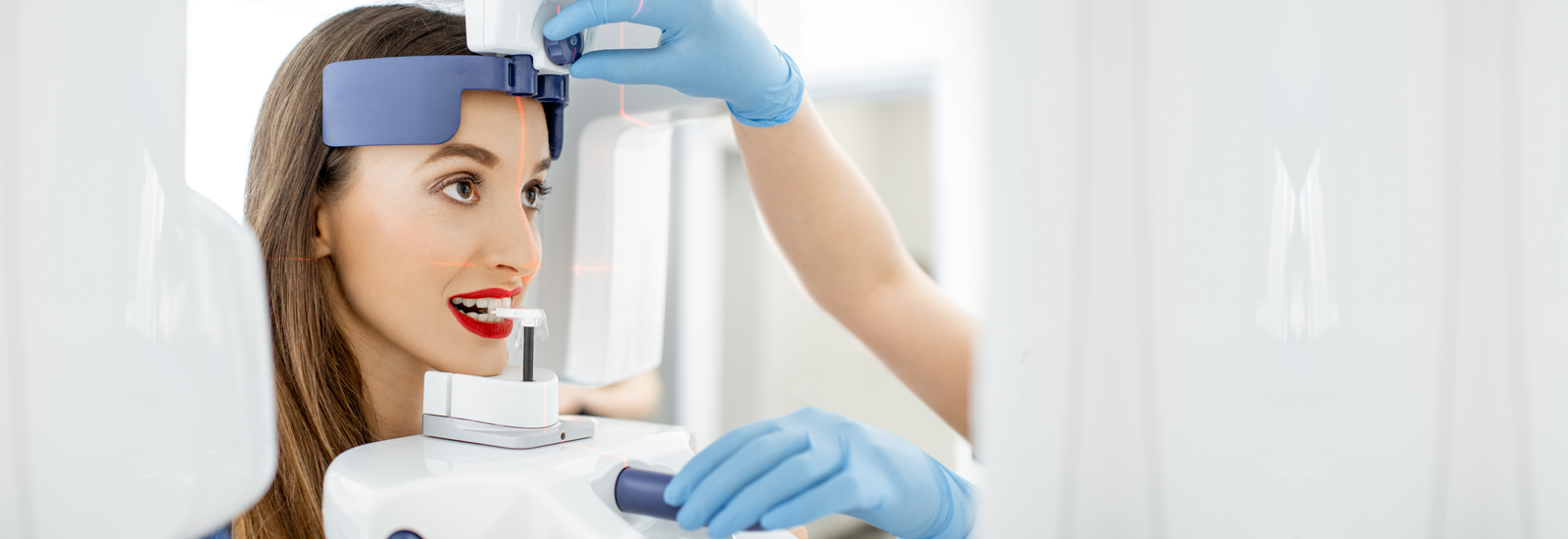
Radiology
Program Objectives
In the Radiography course at California Dental Institute, students will learn the basics of head and neck anatomy to familiarize them with the important anatomical landmarks pertinent to dental radiography. Our students will be taught of radiographic films, digital sensors, radiographic equipment, and most importantly, infection control in the x-ray room. Our course is designed to teach students the basic principles of x-ray radiation and affecting the quality of x-ray imagines and provide hands-on learning of the function and maintenance of the x-ray machines.
Our 32-hour long dental radiography course consists of the following modules:
- Didactic/classroom teaching – 8 hours
- Laboratory training – 12 hours
- Clinical learning and hands-on experience – 12 hours
Learning Outcomes
Upon completion of this course, our students will be able to:
- Identify anatomical landmarks in the head and neck region – the facial and jawbones, nerves and blood vessels, muscles, salivary glands, and jaw joints.
- Understand the uses of radiographic imaging in dentistry
- Learn the discovery of x-radiation
- Learn principles of radiation physics. Understand the properties of x-rays and describe what happens during ionization.
- Identify and learn the functioning of different parts of the dental x-ray machine and the x-ray tube.
- Understand how x-rays are generated
- Learn about types of radiations
- Understand and follow safety principles with regards to dental radiography
- Describe the ALARA (As Low as Reasonably Achievable) principle, including the dentist and dental assistant’s responsibilities concerning dental radiography.
- Describe the long-term biological effects of x-ray radiation on human tissues.
- Learn about the maximum permissible dose (MPD) of ionizing radiation and understand the necessity of monitoring the dental personnel’s x-ray radiation exposure.
- Explain the hazards of primary, secondary, and scatter radiation on human tissues
- Understand the need for operatory radiation shielding, and protection of the patients and staff from radiation exposure
- Name and explain the functioning of the major components of the dental x-ray unit.
- Describe the effect of variables such as the milliamperage, kilovoltage, exposure time, and length of the position indicator device (PID) on the quality and type of x-ray image.
- Explain the need for the last speed film and name the sizes and composition of film used for intraoral dental radiographs.
- Understand the properties of exposed and processed dental radiographic film and the criteria related to obtaining diagnostic-quality radiographs.
- Explain the role of vertical and horizontal angulation for radiographic imaging.
- Independently operate a dental x-ray machine.
- Process different types of dental x-ray films.
- Evaluate radiographs for density, contrast, detail quality, and definition.
- Utilize the paralleling and bisecting techniques in dental radiography.
- Understand quality assurance in dental radiography.
- Learn and process periapical, bite-wing, occlusal, and panoramic dental radiographs.
- Identify the maxillary and mandibular anatomic landmarks routinely evident in radiographs.
- Identify errors – such as overlapping of the image, cone cut, bent film, light radiograph, dark radiograph, fogged film, blurred image, saliva stain, double exposure, herringbone effect, superimposed objects, spots or streaks, static electricity, and scratches on the film – and their troubleshooting.
- Learn appropriate seating and safety measures for dental x-ray patients, observing the universal-precaution and executing infection-control steps.
- Develop and mount radiographs.
Dental Assisting
Radiation Safety (X -ray licence)
8 hours Infection Control
Basic Life Support / CPR for Healthcare Providers
© Copyright California Dental Institute


